|
|
|
Escherichia
coli
Toxin: Watch
What You Eat!
|
|
•Summary |
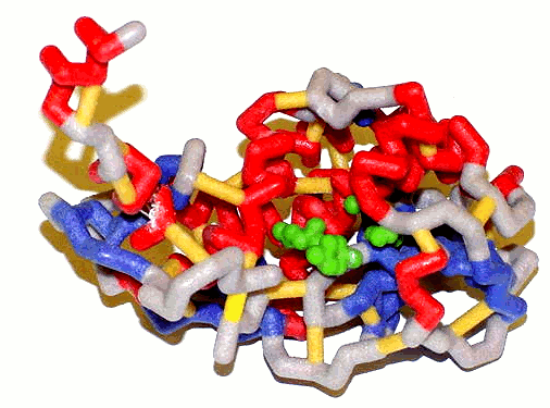
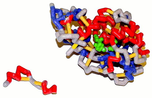 Here is a picture of Subunit A (left), or the active site, of the shiga toxin. On the right, there is E. coli after the glycosidase cleavage.
|
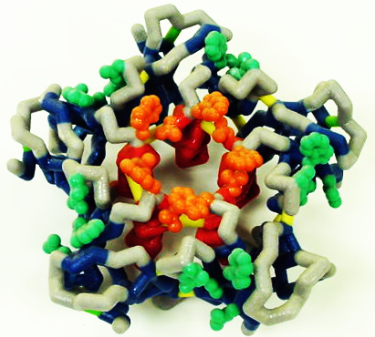 This here is the image of the B subunit, or the binding domain, of the E. coli threat. |
| E. coli uses the following pdb file: 1R4P.pdb |
Background images are from
pdb files downloaded from the Protein Data Bank.
http://www.rcsb.org/pdb/home/home.do
The images are of catalase,
single stranded DNA, and of insulin.
The pdb file numbers
(Brookhaven Extension numbers) are 4BLC, 1EW1, and 2CEU.